Oliveira lab
  • Home
  • Research
  • Publications
    • In the news
  • People
    • Open Positions
  • Gallery
  • Photos
    • CHR 10th Anniversary
    • CHR Official photos
    • CHR Random
    • CHR Moments of Pride
    • CHR Secret Santa 2020
    • Lab Retreat Azores 2018
    • CHR 5th Anniversary
    • CHR 4th Anniversary
    • DrosTuga 2014
    • CHR 2nd anniversary
    • CHR 1st anniversary
  • Contact
  • Knitted chromosomes

WHAT WE DO


During cell division, the genetic information contained in the chromosomes needs to be equally distributed to the new cells that are formed. If the correct distribution of the genetic material is somehow impaired, cells may obtain an abnormal number of chromosomes or even break and loose significant parts of the genome. These abnormalities are usually associated with many health conditions, such as cancer development, genetic disorders and infertility. Our lab investigates how chromosomes are assembled and how their morphology influences the fidelity of cell division. 

We are focused on three main questions:

  • How are chromosomes assembled during nuclear division?
  • How does chromosome structure influence the fidelity of mitosis?
  • How do failures in mitosis impact on development and tissue homeostasis?


OUR APPROACH


Picture
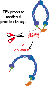
We use a multidisciplinary approach combining acute combining acute protein inactivation, 3D-live cell imaging and quantitative methods to probe for chromosome morphology and mitosis progression (see gallery). Our "acute approach" relies on a major technical advance that enables specific, rapid and efficient protein inactivation in a tissue- and/or time-dependent manner (see Pauli et al Dev Cell 2008 and Oliveira et al NCB 2010). This method uses an exogenous protease (Tobacco Etch Virus, TEV) to cleave an engineered protein of interest that contains TEV cleavage sites. 

Picture
  • Why flies?
We use Drosophila melanogaster as a model system. Fruit flies are an excellent model to address chromosome structure, as they offer a powerful system for simple genetic manipulations combined with live cell imaging of many mitotic tissues in which condensed mitotic chromosomes can be easily visualised. Additionally, they offer the unique opportunity to study the consequences of aberrant chromosome structure in the context of a developing organism. 

PUBLICATIONS

IMAGE GALLERY

IN THE NEWS


COLLABORATORS

  • Béla Novák, Department of Biochemistry, University of Oxford, UK
  • Bill Sullivan, Molecular, Cell and Developmental Biology, University of California

FUNDING

Picture
Picture
Picture
Picture
Picture
@IGC
Picture
© 2022 Raquel Oliveira
  • Home
  • Research
  • Publications
    • In the news
  • People
    • Open Positions
  • Gallery
  • Photos
    • CHR 10th Anniversary
    • CHR Official photos
    • CHR Random
    • CHR Moments of Pride
    • CHR Secret Santa 2020
    • Lab Retreat Azores 2018
    • CHR 5th Anniversary
    • CHR 4th Anniversary
    • DrosTuga 2014
    • CHR 2nd anniversary
    • CHR 1st anniversary
  • Contact
  • Knitted chromosomes